Understanding the Anatomy of a Horse’s Hoof
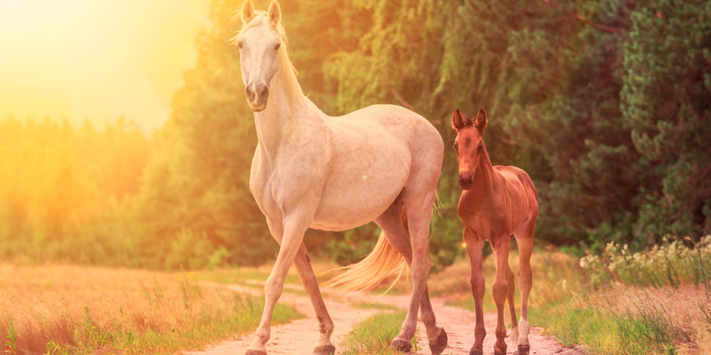
When you take into consideration the size of a horse relative to the size of a hoof, it’s easy to see what an amazing engineer Mother Nature is.
I have always been told, “no hoof, no horse.”

Because of this it is extremely important for us as owners to understand the anatomy of the equine hoof so that we are able to keep our horses as healthy and sound as possible.
Hooves may look simple but in reality they are extremely complex structures.
The anatomy of the equine hoof can be intimidating, but the hoof can be broken down into three groups to make it easier to understand.
Anatomy of a Horse’s Hoof
Inner Structures
Digital Cushion
The digital cushion is a mass of flexible material that lies below the coffin bone.
It contributes to the formation of the heels and acts as one of the primary shock absorbers in the hoof.
This area of the hoof becomes compromised in horses with a long toe, low heel conformation.
This type of conformation causes the heels to bear more weight than normal which can slowly compress the cushion.
Once this occurs, the digital cushion will not regenerate.
Collateral Cartilages
The collateral cartilages are located at either side of the coffin bone and extend upward.
Their purpose is to help the hoof expand when bearing weight.
Hardening of these can cause limitations in the hoof’s natural expansion which can result in some sensitivity when checked with hoof testers.
This is most commonly an issue in heavy breeds who are frequently worked on hard ground.
Coffin Bone
The coffin bone is the outer hoof capsule’s anchor.
It has a large, porous surface that is ideal for soft tissue attachment.
Anything that interferes with the working relationship between the coffin bone and the hoof capsule can result in lameness in the horse.
The coffin bone can also serve as a significant indicator of equine hoof health through examination of the palmar angle (between the bottom of the coffin bone and the ground).
A normal palmar angle is slightly positive, which would mean conformationally a horse has heels slightly higher than the toe.
If the angle would be too positive, like greater than 10 degrees, there may be a club foot or laminitis at play.
A zero or negative palmar angle would indicate a horse has heels lower than the toe.
This is the kind of scenario that would result in a compromised digital cushion as described above.
Navicular Bone
The navicular bone is a small bone that helps to stabilize the coffin bone.
This also anchors the deep digital flexor tendon, which runs down the back of the leg and allows the leg to bend and flex.
The navicular bone undergoes significant compressive force due to this tendon.
This bone is associated with navicular disease, which is a common cause of lameness in horses.
Outer Structures
Hoof Wall
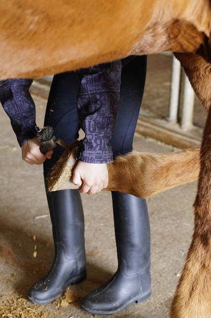
The part everyone sees, the hoof wall is a continuous growing, keratinous material that needs to be worn or trimmed off periodically.
There are no blood vessels or nerves in the hoof wall.
This hard outer covering protects the delicate structures within it, supports the weight of the horse, and absorbs the shock associated with movement.
Healthy hoof wall should not have any cracks or rings.
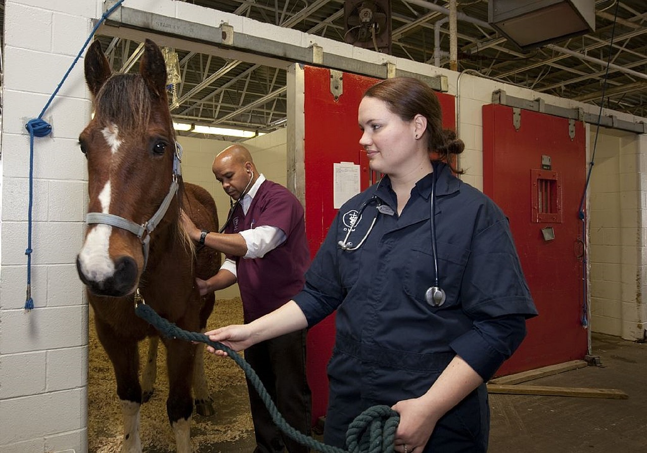
Rings can indicate an underlying issue in the horse’s health that is affecting the hooves.
A veterinarian should be consulted.
Cracks can expose internal structures to potentially damaging environments, which can lead to lameness.
Coronary Band
The coronary band is the area at the very top of the hoof wall where the hoof meets the hairline.
This is where new hoof growth is generated from.
A healthy hoof should grow at about a rate of ⅜ inch per month.
Although this area is structurally strong, it also contains a large blood supply to support hoof growth.
An injury to the coronary band can result in damage to the hoof wall or even cause permanent issues with hoof growth to the point that the horse may be deemed unrideable.
Periople
The periople on a horse is similar to the cuticle of a human.
It is a waxy substance that covers the outside of the hoof wall for added protection and to prevent excess drying.
Corium
What anchors the coffin bone to the hoof wall is the corium.
The corium is made up of soft tissues including blood vessels, nerves, and the laminae.
Laminae are interlocking leaf-like structures that are primarily responsible for attaching the hoof wall to the coffin bone.
These laminae bear the majority of the weight of the horse.
If this structure was damaged, and the bone to hoof wall attachment disrupted, the corium can start to deteriorate which can lead to the coffin bone dropping in the front or completely sinking.
Bottom of the Hoof
Sole
The sole is the bottom side of the hoof.
Due to the slight concavity of the equine hoof, the sole very rarely comes into contact with the ground.
The structure of the sole is very similar to that of the hoof wall, with the only exception being the sole is slightly softer than the hoof wall.
A very important part of the sole is the white line, which can actually be slightly yellowish in color.
This is the area where the hoof wall and sole merge.
If this area of the sole becomes impaired, germs may enter into the hoof and work at separating the hoof wall layers.
Once this has occurred, these germs can spread throughout the entire hoof and can lead to lameness in the horse.
Frog
The frog is a highly elastic, wedge shaped mass on the bottom of the horse’s foot that is usually the first to make contact with the ground.
It provides protection to the digital cushion beneath it, assists in traction and circulation in the hoof, and acts as a shock absorbed when the horse moves.
As the horse moves, the digital cushion and frog act together as a sort of pump, circulating blood through the hoof.
This is why exercise is necessary for generating hoof growth.
Bars
The bars are a continuation of the hoof wall and serve as braces to keep the heels from contracting.
This area also contributes to building the sole of the hoof and helps to support the horse’s weight.
Central Sulcus
There are grooves down the center and either side of the frog.
The center groove is known as the central sulcus, while the grooves down either side are known as the central and lateral sulci.
The central sulcus should be fairly wide and shallow.
In cases where it is narrow and deep, the horse may have contracted hooves or sheared heels.
These narrow and deep central sulcus can harbor moisture and germs which can in turn lead to thrush.
We should always be caring for our horses’ hooves with regular trims, a healthy diet, and plenty of exercise, and always be sure to watch out for any changes in the outer structure of the hoof.
It is much easier (and usually cheaper) to prevent issues rather than treat them once they have already occurred.
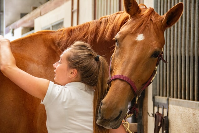
We all try our best to keep our horses as healthy and happy as possible, and for many of us hoof health is easily forgotten.
But, always remember, “no hoof, no horse.”
Interested in learning more about the equine hoof? Check out The Humble Hoof podcast to feed your newfound interest in hooves!

Lauren is an internationally published author, trainer, and has helped hundreds of horse-rider combinations create lasting bonds and the success they desire. Check out Lauren’s incredible story: From horse-crazy girl to international equine educator. Or if you want to send Lauren a quick message, check out her contact page here.